Glioblastoma multiforme is the most aggressive brain tumor and it’s also the most frequent one. Depending on where it grows inside the brain, it can cause plenty of different symptoms, from a simple headache to movement impairment.
Table of Content
What are the different types of brain tumors
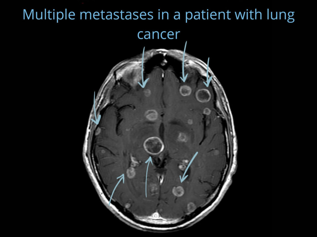
There are many different brain tumors, and each of them has different behaviors. There are malignant tumors and benign tumors. Some are slow-growing, others are aggressive. There are tumors that “are born” inside the brain and others that “travel there” from another organ (metastases).
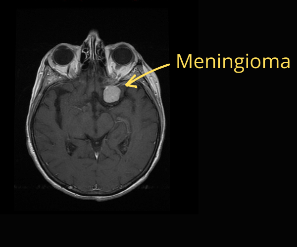
Some tumors grow by “pushing” structures around them and others infiltrate those structures, making them very difficult to remove. For example, if you have a small meningioma (a benign tumor) you may never have any symptoms. Sometimes, it won’t even need treatment.
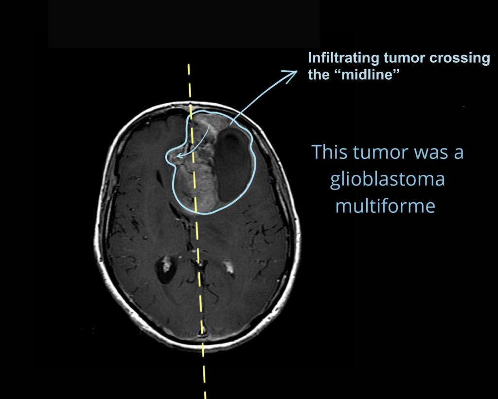
But if you get advanced glioblastoma multiforme, that one grows pretty fast, so you are going to get symptoms quickly, and you’ll need treatment to increase your survival.
What is glioblastoma multiforme?
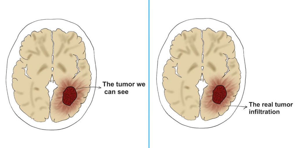
Glioblastoma multiforme is a primary brain tumor. That means it is “born” inside the brain (as opposed to metastasis). It belongs to a group of tumors called astrocytomas, and glioblastoma is the most aggressive amongst them. It grows very fast and infiltrates the structures around it. That makes it hard to remove it completely because there are always some malignant cells remaining in the tissues around it.
Unfortunately, not only it is aggressive, but it is also the most common primary malignant tumor in the brain.
What are the symptoms of glioblastoma multiforme?
Pick any neurological symptoms you can think of: glioblastoma can cause them. Our brain is a complex organized structure: each part of the brain cortex is responsible for a certain function: moving, taste, hearing, memories…
Depending on what part of the brain is affected, the symptoms will vary. If the tumor grows in the area for “left-hand movement” you’ll have trouble moving your hand. If it grows in the “speaking area”, you’ll have aphasia (it means “unable to speak”). And so on…
It can also cause headache and other unspecific symptoms. However, most people with a headache don’t have a brain tumor, they just have a headache. Headache becomes more worrying if:
- the pain is worse during the night (it wakes you up)
- if there are changes in personality
- and especially if you have other focal symptoms (you can’t move your arm, leg, can’t see…)
If you have some of these alarm symptoms, go to your doctor.
How do you diagnose glioblastoma multiforme?
First of all: if you have any of the above symptoms, go to your doctor. They will examine you, checking one by one all your neurological functions. After that, they will get a first impression to decide whether you need further tests.
C.T.
The first test you get is usually a C.T. It is accessible and fast, and it usually tells us if there is something wrong in the brain. If you have a brain tumor, C.T. will show it in most cases.
MRI
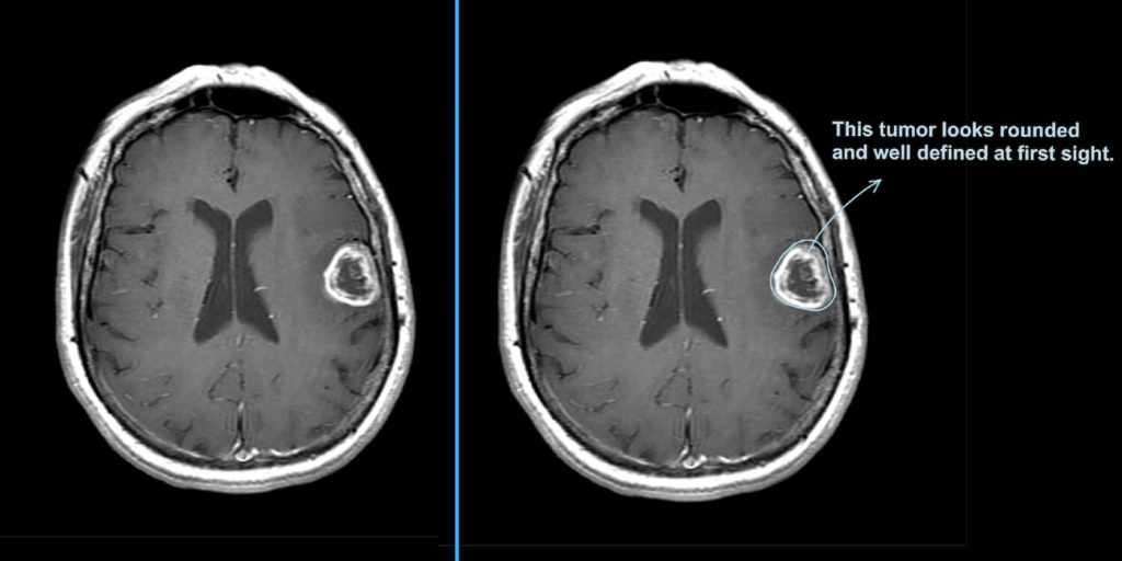
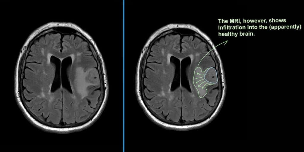
Then, they’ll usually admit you to the hospital to give you some medication (to decrease the inflammation around the tumor) and to have an MRI. MRI shows the tumor in greater detail.
To really make sure what type of tumor it is, the neurosurgeon must take a biopsy of the tumor, a sample. After analyzing that sample, they can confirm whether it’s glioblastoma multiforme.
How do you treat glioblastoma multiforme?
We usually treat it through a mix of techniques:
Surgery + Radiotherapy + Chemotherapy
Surgery
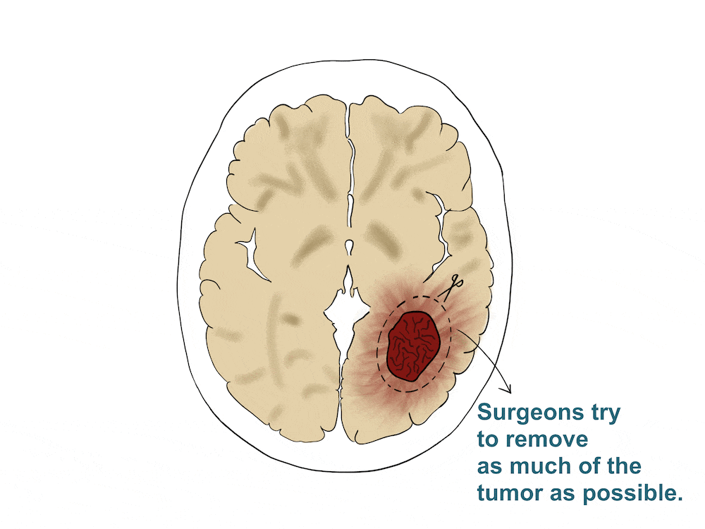
First, neurosurgeons will try to remove as much of the tumor as possible (maximal surgical resection). However, there are some limitations. First, the tumor is sometimes infiltrating crucial brain areas, which we must not touch. For example, imagine a tumor is infiltrating your “breathing center”. We can’t remove that safely because if we damage the breathing center the patient will die.
Second, glioblastoma multiforme is a very infiltrating tumor. So, you may see “a ball”, but in reality, there are thousands of malignant cells all around it, in the apparently healthy tissue. That’s why surgeons try and remove as much “healthy” tissue as possible around the tumor. Sometimes, it’s just impossible to remove the whole tumor.
Radiotherapy
After surgery, most patients receive radiotherapy. The goal? To kill those cells that are still remaining in the surrounding tissue.
Chemotherapy
We use chemotherapy to remove any malignant cells left. The most common agent is Temozolomide, which is specific for glioblastoma multiforme. It comes in a capsule that you take once a day.
With these treatments combined, the average survival is 10-16 months. Fortunately, there have been some new advances in recent years.
Other therapies for glioblastoma multiforme:
Gamma-knife (Stereotactic Radiosurgery)
The name is misleading because it’s not a surgery. It’s more like radiotherapy. With gamma-knife, the radiotherapy beam is so focused that its effect is like that of a knife’s. Like a laser. It “burns” the tissue. Also, the kind of radiation that is used releases a lot of energy on a specific point, but very little around it. That’s great because we can direct the beam towards the tumor, and we won’t damage the surrounding structures.
You will need to wear a special thermoplastic mask (or a stereotactic frame), which are custom-made for you. They will provide you with it in the hospital and you have to wear it during your gamma-knife sessions. Doctors will obtain an MRI of your brain with your mask on. That way, they will know the exact “coordinates” of the tumor, using the mask as a reference. Then, they’ll use those coordinates to aim at the tumor.
Awake surgery
Awake surgery is a procedure where the surgeon performs surgery on you while you are awake. This is possible because they apply local anesthesia to your scalp, so they can cut through it.
Surprisingly, your brain can’t feel any pain or sensation when they touch it or cut it.
Awake surgery is only performed at very specialized centers. Neurosurgeons use it for tumors that are too close to functional areas. For example, imagine the tumor is close to the speech area. If they “cut too much” the patient can lose the ability to speak. But they still want to remove as much of the tumor as possible. The way to obtain a good balance between removing “too much” or “too little” is awake surgery.
With awake surgery, surgeons open the patient’s skull. When the brain and the tumor are exposed, they wake the patient up from sedation. Then, they ask the patient to do some things, like speak, add some numbers up, etc. Meanwhile, they are looking at the brain and checking which areas are “lighting up”. That way they know the areas they’ll have to preserve.
Wafers
Chemotherapy wafers are small implants that contain some chemotherapy inside. They are left in the cavity after they remove the brain tumor, during the same surgery. The most common are Gliadel® wafers, which contain carmustine inside (a chemotherapy agent).
As the wafers start to degrade, they release carmustine to the tissue around them, killing the surrounding malignant cells.
Tumor treating fields
Scientists first discovered this therapy in 2004. They found out that if they applied electric fields to a tumor, it slowed down growing. Tumor treating fields consist of a power source, field generator and a transducer arrays. It’s a non-invasive treatment. Patients have to wear a helmet with the transducer arrays virtually all the time (they can unplug it for showers) and they also have to shave their head.
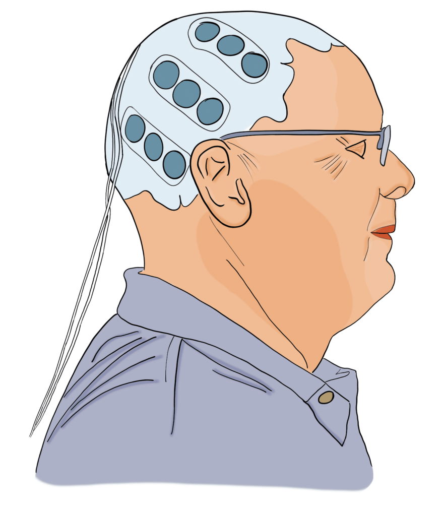
Then, the arrays deliver low intensity, alternating electric fields through the skin. These fields slow down tumor growth and can increase overall survival by 5 months. Their most frequent side effect is mild cutaneous irritation.
You can read more about it here:
Leave a Reply