The femur is the strongest bone in your body, so it usually takes a powerful hit to break it, like a car accident. Unlike hip fractures, which happen in old people who fall, femoral shaft fractures tend to happen in young people who suffer high-energy accidents. This article, is about them, femoral shaft fractures. If you want to read about hip fractures, click here.
3 Different Types Of Femoral Fractures
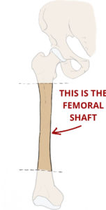
There are 3 different parts of the femur that can break:
· The proximal femur: it’s the upper part, the hip.
· Shaft fractures: the long part in the middle. This is the type of fracture we’ll be talking about in this article.
· Distal femur: the lower end, part of the knee.
How do you break your femur?
People who break their femur are usually young people that are involved in a high-energy accident (car/motorbike crash, big fall…). That’s because the shaft is really tough, and it takes a powerful trauma to break it. The distal femur is tough to break too, and it usually happens during sports accidents.
Another cause for femoral fractures are pathological fractures. They happen in a bone that has a lesion inside, usually metastasis. The lesion weakens the bone structure, making it prone to break.
5 Things That Can Go Wrong (Complications)
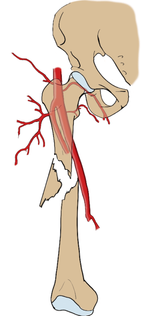
When you break your femur there is a life-threatening complication: hemorrhage. During such a traumatic hit, the fragments of femur can cut through one of the arteries that run next to it, and cause a large bleeding. What’s worse, it can go undetected as it will be an internal bleeding, inside your thigh (unless it is an open fracture).
If the fragments move a lot, they can pierce through the skin, and you can see some bone coming out. It is really painful of course, but it also increases the risk for infection. If the wound gets dirty with mud, dust… then chances for infection will skyrocket. The first thing to do with an open fracture is cleanse it with physiological saline and then give the patient plenty of antibiotics.
Also, your femur can break with a “clean” fracture or in multiple pieces. If that’s the case we call it a comminuted fracture, which is bad news because it makes it hard for us to stabilize the fracture and make fragments stick together.
Another dangerous complication is fat embolism: inside every long bone (like the femur) there is medullary fat. When the femur breaks, some fat can enter the bloodstream and travel to some other organs. When that happens, small arteries can be obstructed, which can be devastating if it affects the kidneys, lungs or brain.
Compartment syndrome: when a person breaks their femur, the soft tissues around the femur suffer as well. The muscles, ligaments… get bruised and swollen. Sometimes, this increases the pressure in their leg enormously. So much that their veins will start to collapse. This, in turn, will increase even more the swelling and pressure. If pressure keeps increasing, arteries will finally start to collapse, causing extreme pain and endangering the leg.
How do we diagnose femoral fractures?
Over 90% of them can be diagnosed with an x-ray, which will show the displaced fragments.
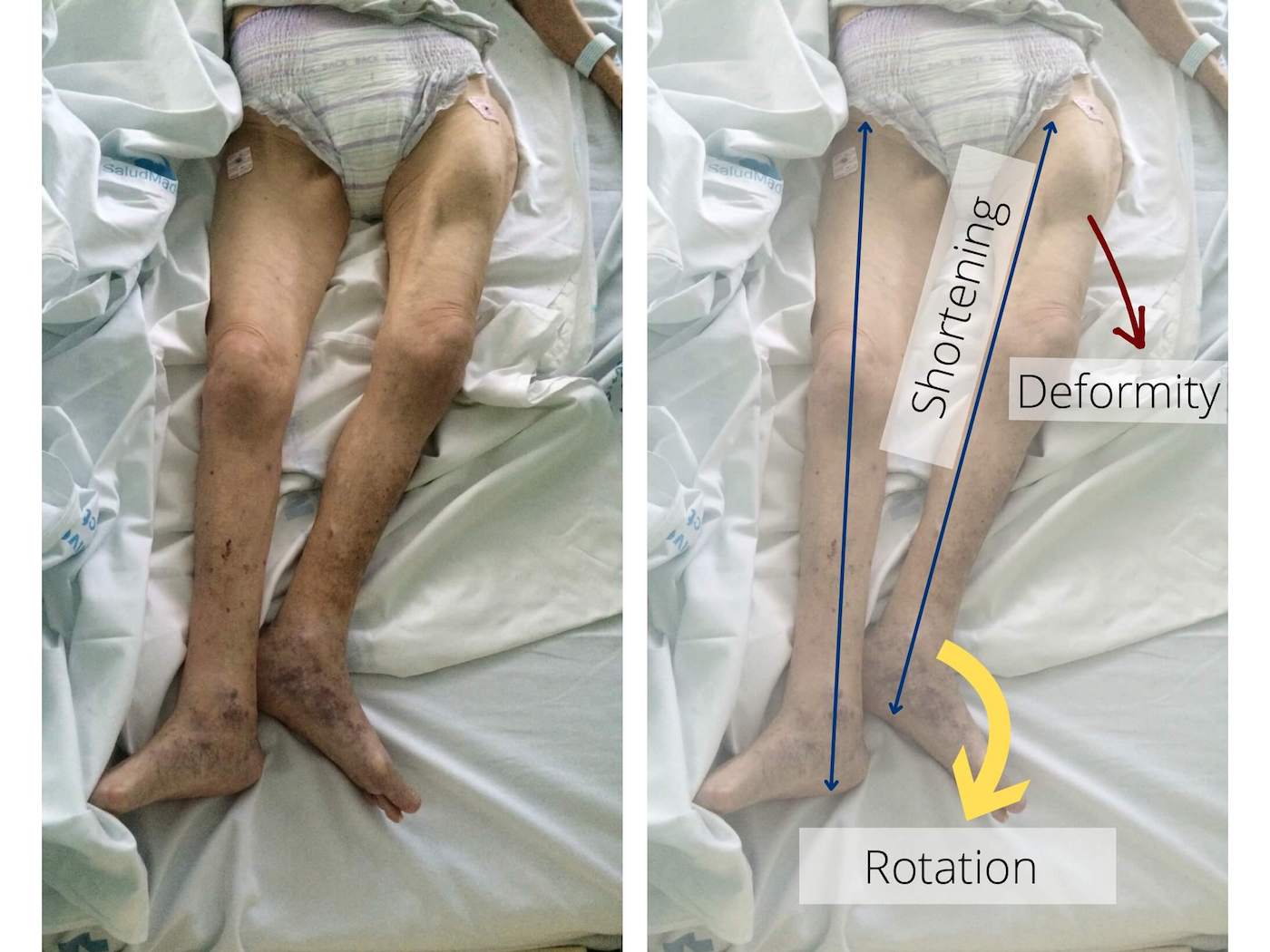
The typical femoral fracture will show the following signs:
- the patient had an accident
- has tremendous pain on his leg
- his leg is shortened
- can’t walk or even stand on his leg
- has a deformity, with the foot typically rotated outwards
If there are any doubts by looking at the x-ray, then we can perform a C.T. scan. It will show better detail on what the fracture looks like, how many fragments there are, and help with surgical planification.
How do we treat femoral fractures?
Immediately after admission
If you have an open fracture, the first step is to cleanse it with physiological saline. Later, during surgery, doctors will wash it with soap or povidone iodine (under anesthesia). Also, in the case of open fractures, they will give you i.v. antibiotics to prevent infection.
Then you will go to the radiology room to get an x-ray. This will confirm the diagnosis and show the fracture type.
Traction: in order to stabilize the fracture and control the bleeding you may need traction. Traction seems a like bit primitive when you first see it: they tie some hanging weight to your leg through a pulley. That way both fragments will get realigned so that when they take you to the operating theater it will be easier to insert the nail.
If there are other body injuries that require urgent treatment, they may give those lesions priority. For example, brain bleeding, which might need urgent neurosurgery intervention.
Once the patient is out of danger
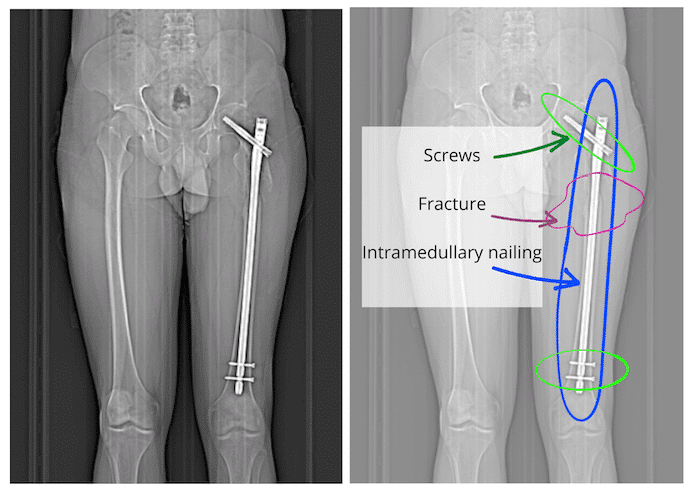
A broken femur needs surgery. Most of the times, it requires intramedullary nailing. If it’s not possible to perform definite surgery in 24-48 h, then external fixation can be a provisional solution. It is used very often.
External fixation
It consists of a metal bar screwed to both fragments of the femur through the skin. Thus, the bar keeps the fragments close to each other and aligned.
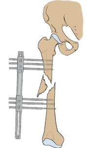
In order to place it, the orthopedic surgeon has to make small cuts on the skin and drill the bone. Of course, this is also done under anesthesia.

This is an external fixation for both ankles (not for the femur), but it shows the idea.
After that, you will need intramedullary nailing: a long nail that is placed inside the femur, crossing through the fracture site and keeping the fragments together and aligned. Then, some short screws are placed on both sides of the nail, to prevent it from moving. One of the benefits of intramedullary nailing is that it will allow you to stand up and walk soon after surgery.
What to expect afterwards
Intramedullary nailing is a very effective technique. It allows patients to start walking soon after surgery. Actually, it’s important that you walk, because as you start walking, the weight on the fracture will increase pressure between both fragments. This pressure works as a stimulus for bone growth and callus formation.
Some drugs you may need after femoral fractures
- Antibiotics: specially if you had an open fracture. They will usually give them to you via i.v.
- Anticoagulant: while you are not walking they will give you some anticoagulant to prevent clot formation.
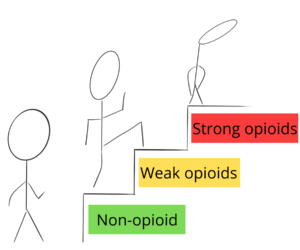
- Painkillers: you should take those that are enough control your pain, avoiding if possible strong opioids.
When things don’t go as planned (Sequelae)
- Shortening of the leg: your leg can get slightly shorter than the healthy one. In most of the cases, the difference is minimum and won’t cause any trouble in the future. If there is a difference of more than 2 cm then you may need to use some custom made shoes (where one is higher than the other).
- Pseudoarthrosis: if there isn’t enough pressure on bone fragments then a bone callus may never form. That is called pseudoarthrosis: instead of the fragments fusing together, a space remains between them, giving it the appearance of a new joint.
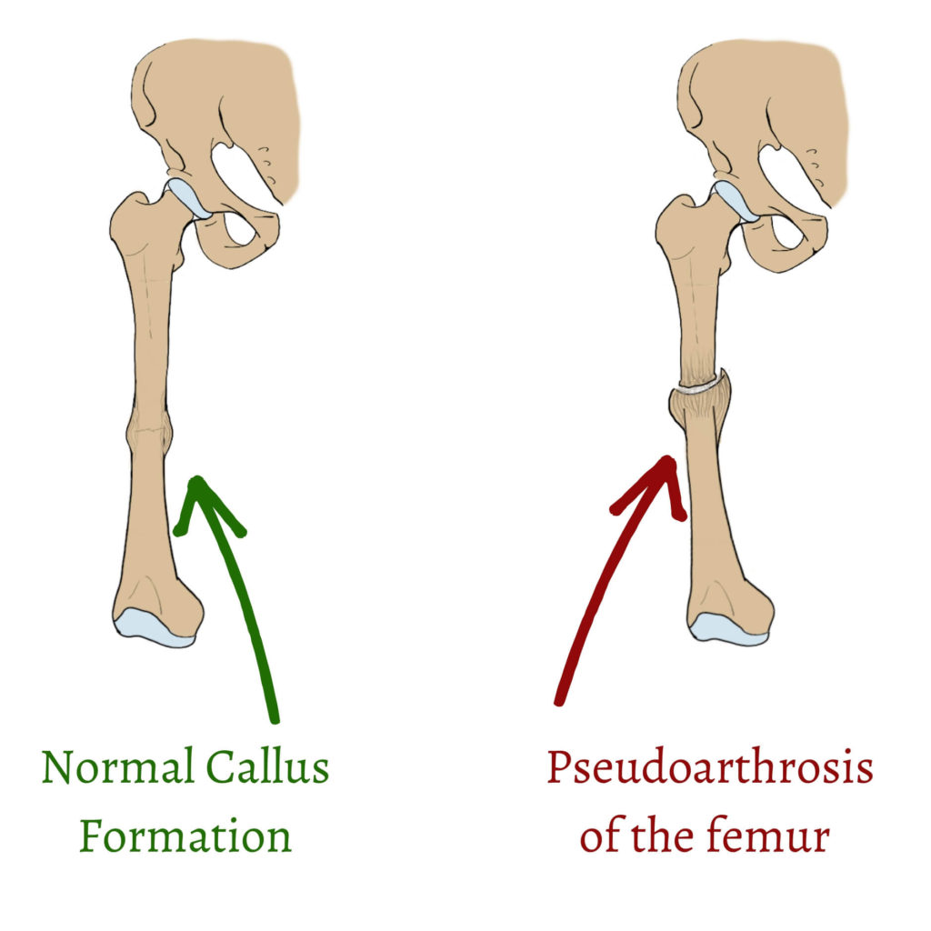
Pseudo = “kind of”; arthros = “joint”; pseudoarthrosis = “kind of a joint”
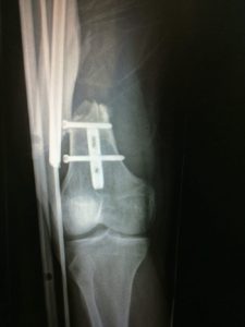
- Myositis ossificans: it’s an abnormal calcification in tissues other than the bone, in the muscles in this case. It usually happens after trauma, like this example.
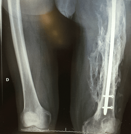
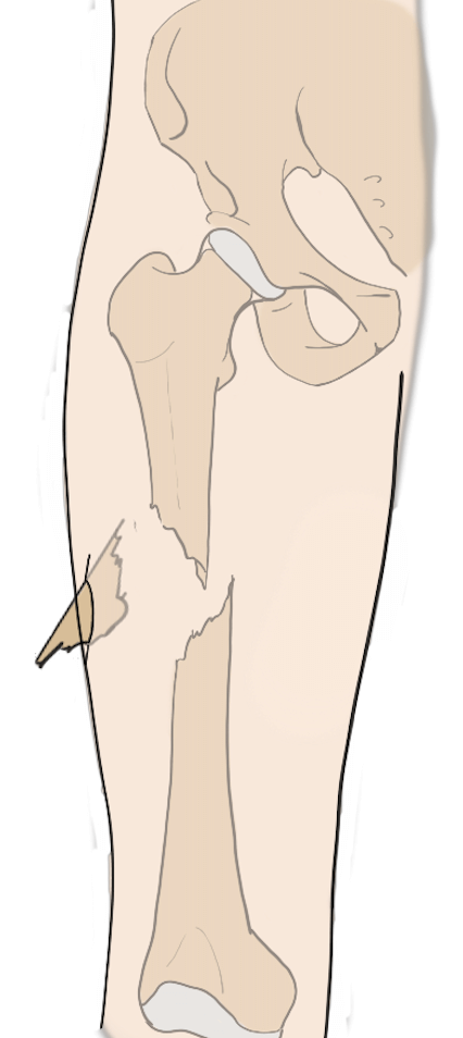
- Peri-implant fracture: once the nail is placed inside the femur, it usually stays there forever. However, the nail can overload the femur, so that there can be new fractures around the nail. Sometimes, even the nail can break, as you can see in this x-ray.
The Rehab Process
In the first 48h, if everything went alright, you will start standing up and even taking some small steps with the help of a therapist and crutches. The physical therapist will teach you how to move safely, reducing the risk for falls and new lesions.
It’s important to start walking soon because that’s when callus formation starts. If you don’t make the effort, the fragments may never fuse. Your physical therapist will also help you with passive movements. They will move your leg and flex/extend your knee so that you don’t lose range of motion. You won’t be able to move your leg at the beginning, so it’s important that some therapist helps you so that your joints won’t get stiff. Then, you will progress from there, first with both crutches, then with only one and, with time, by yourself.
All your progress must be smooth and progressive, and you must always follow your surgeon’s advice. Not all surgeries are the same and neither are all patients.
Questions And Answers
-
Can a broken femur heal on its own?
No, not a femoral shaft fracture. As we explained a femoral shaft fracture is a pretty bad injury. You won’t be able to even move your leg.
-
How long does it take to recover from a femoral fracture?
It depends on each case, of course. We can say between 3 and 6 months, as a general estimation.
-
-
Is a broken femur dangerous?
-
Yes, it can bleed a lot, endangering your life. Also, it can get infected if it’s an open fracture. If it doesn’t heal correctly it may require several surgeries… so, yes, it is dangerous.
Leave a Reply